Cranial Nerves Definition
The fibers of the cranial nerves with motor function (efferent) originate from cell groups that are found deep.
Firstly in the brain stem (motor nuclei) and are homologous to the cells of the anterior horn of the spinal cord.
Secondary fibbers from the cranial nerves with sensory or sensory (afferent) function have their cells of origin (first-order nuclei) outside the brain stem.
Usually in ganglia that are homologous to those of the dorsal root of the spinal nerves.
And also Second-order sensory nuclei are founding in the brain stem—functional classification.
What are Cranial Nerves functional aspect.
- Firstly I, II, and VIII are keening to particular sensory input.
- Secondary III, IV, and VI control eye movements, photometer reflexes, and accommodation.
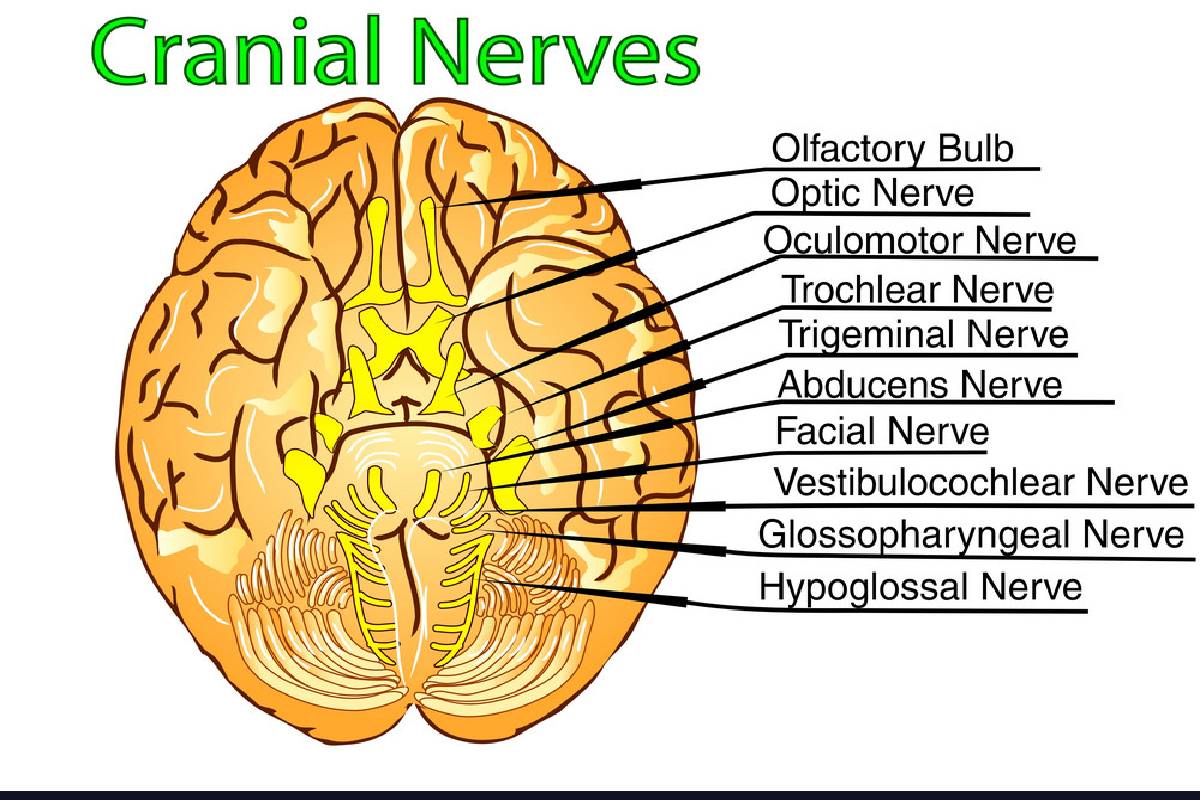
What are the characteristics of each cranial nerves?
1. Cranial nerve: olfactory nerve
- Cranial nerves is a sensory nerve that gives rise to the sense of smell.
- The cranial nerves fibers originate in the bipolar cells of the olfactory mucous or yellow spot, located in the upper portion of the nostrils.
- These cells possess ascending axons that constitute the olfactory nerve fillets and also the nerve fibers from the bipolar cells.
- which cross the lamina of the ethmoid and also reach the lower face of the olfactory bulb.
- In close contact with the cranial nerves, are a small pair of nerves called terminal nerves.
2.Cranial nerve: optic nerve
- It is a sensory nerve that emerges from the eyeball. And also the nerve that allows us to see.
- It originates from the ganglion cell layer of the retina. And also it is the anterior angle of the optic chasm.
- Route and relationships: this nerve measures approximately 4 cm.
- However in length, it goes up, back and in.
- Four segments are labeling in its first segment intramuscular.
- The axons of the tumor cells of the retina converge at the optic after here, and the nerve pierces the superficial layers of the eye (sclera and choroid) at a site called the zonal.
3.Cranial nerve :Terminal branches
- However upper terminal branch Innervates the superior rectus muscle of the eye and the superior elevator eyelid.
- The lower terminal branch innervates the internal rectum, and also the lesser oblique, and the lower rectum.
What is Cranial nerve: pathetic nerve or trochlear nerve?
- It is the single cranial nerves that crosses its fibers inside the brain stem.However it is an exclusively motor cranial nerves that supplies only the more significant oblique muscle of the eye.
- Therefor fibers that come from this core, more over playing on the surface, intersect with those on the opposite side.
- It is the only nerve that emerges from the posterior aspect of the brain stem in cranial nerves.
- Ans also, It emerges on the posterior aspect of the cerebral peduncles, on each side of the valve pendulum of Viennese’s.
- It penetrates the external wall of the sinus and is located initially below the universal ocular motor and above the ophthalmic engine.
- And also it contains the lateral faces of the cerebral peduncles and goes forward, the direction of the cavernous sinus.
- Usually enters the orbit through the spheroid cleft and usually passes outside the ring of Zion in cranial nerve.
- Its terminal branch penetrates the more significant oblique muscle of the eye, which is usually innervates.
What is Cranial nerve: trigeminal nerve?
- It receives the sensitivity of the integuments however the anterior two-thirds of the skull, of the entire face, nostrils, orbital, oral cavity, and its contents.
- In turn, it is the motor nerve of the chewing muscles and also some others.
- Sensory fibers of this nerve are born in the ganglion of Gasser, located at the apex of the supererogation however the aspect of temporal rock. Said ganglion has an upper face, a lower front, a convex interpolate edge, and a concave, posterior inner edge.
- The interpolate border of the Gasser’s ganglion gives rise to fibers that constitute the ophthalmic nerves, the upper maxilla, and the sensitive part of the lower maxillary nerve.
Also Read : What is CBD oil?-Definition, Benefits of CBD oil and MoreWhat is CBD oil?-Definition, Benefits of CBD oil and More
- READ MORE:- healthyseo01